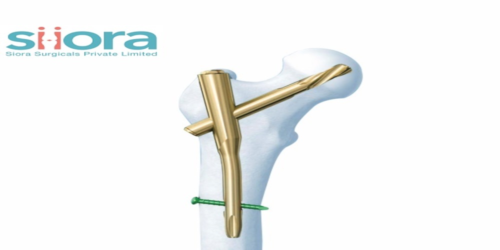
There are many devices indicated for intramedullary fixation and several different techniques for their application. Orthopedic Implants Manufacturing company in India use an advanced technology to manufacture implants and surgical instruments. Intramedullary devices can be hollow or solid; triangular, circular; or cloverleaf in cross-section; and flexible to extremely rigid. They can be placed in either undreamed or reamed channels, can be used with or without interlocking screws, and confer different degrees of fixation stability to the fracture. The intramedullary nail can be inserted without exposing or opening the fracture site. The nail is considered a load-sharing device because the central position of the nail allows the bone to carry some of the load. Intramedullary nails are used most commonly to treat fractures of the tibia, femoral and humeral shafts.
The terms nail, rod, and pin have specific biochemical implications, but clinically they are used interchangeably and often represent the original name of the device (for example, Rush pin, Sampson rod, Ender nails, Kuntscher nail). Several early nails were solid and designed to be driven into the bone without reaming (for example, diamond-shaped-Hansen-Street nail). The term nail was usually used for intramedullary devices that were driven into reamed or unreamed channels. They are, in a sense, wedged position, which makes them self-retaining. Some of these nails are still being used. A rod was a hollow or solid intramedullary device with a blunt tip that was larger than that of a nail and that was usually placed in reamed channels. Intramedullary pins are solid, smaller, circular in cross-section, and usually driven into the canal without reaming. Nails, rods, and to a lesser degree-pins neutralize shearing and bending forces but allow rotation and axial compression.
Most intramedullary nails and rods used now are hollow, and almost all have a unique cross-sectional pattern designed to prevent or reduce rotation. The hollow designs have either a closed or an open section. They provide stability by an interference fit along the medullary canal. However, the success and availability of interlocking techniques have made most surgeons reluctant to depend on only an interference fit to provide stability for several types of fractures. Many intramedullary devices are quite the same, with only small differences in design that may not be radiographically apparent, making identification difficult.
The Rush intramedullary pin may be used in fractures of the smaller tubular long bones. One end is beveled for placement in undreamed channels; the other end is hooked, which facilitates removal and prevents migration. Ender’s nails are oval in cross-section, solid, and flexible. They were originally developed for femoral fractures, including femoral neck and intertrochanteric fractures. Usually, multiple nails are driven into the medullary canal through drill holes at the end of the bone. They are less able to resist axial and rotational loading forces. The Sampson intramedullary rod is a thick-walled, very rigid that is slightly curved and has a fluted surface to prevent rotation. It is used primarily in re-constructive processes, such as knee arthrodesis. Kuntscher nails are cloverleaf in cross-section and hollow, with an open section and a rounded tip, allowing for a press fit.

